When scientists perform experiments in the lab, they require models that mimic, at least in part, the subject of study, which could be a developmental process, a disease, or other phenomena. These models can be broadly divided into two main categories: “in vitro” and “in vivo.” Though the definitions may vary among scientists and depend on the subject of study, generally, an “in vivo” model refers to the use of a living subject, such as an animal, a plant, or even yeast. In contrast, an “in vitro” model uses parts of the living subject of interest. For example, cells originally from a specific organ of an animal, like epithelial cells from the intestine, can be used. These cells can be obtained from tissue samples taken from that organ. For instance, a patient’s biopsy can be used to produce what is known as a stable or immortalized cell line.
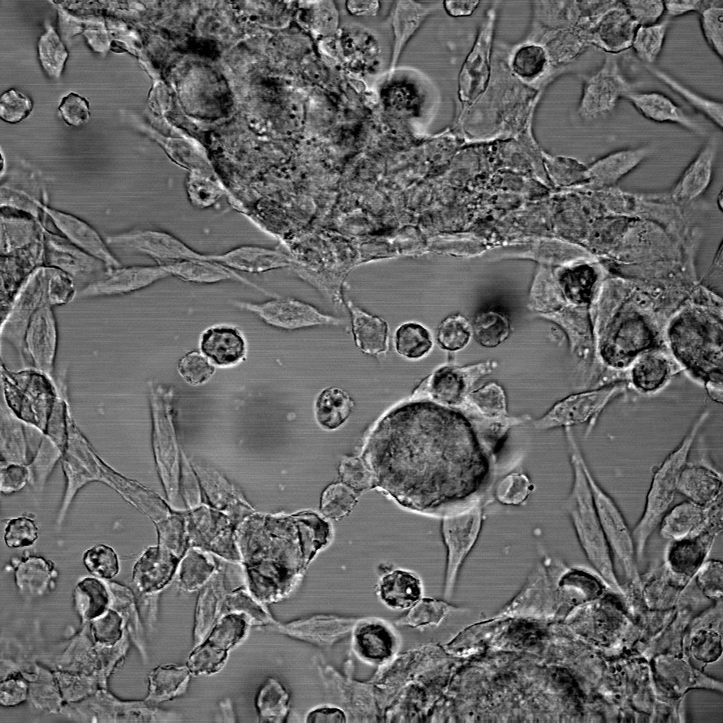
Briefly, the tissue fragment is taken to the lab and disaggregated to obtain a cell suspension, where cells lose their connections to each other and to the extracellular components or matrix. These cells can then be grown in a special plastic container with a specific growth medium that provides all the necessary nutrients for survival. Adherent cells, like epithelial cells, will grow by attaching themselves to the plastic surface, forming a layer of cells. This type of in vitro cell culture is called 2D (two-dimensional) because cells grow in two directions. Normally, they include only one cell type, though modern culturing systems allow for the co-culture of two cell types to study their interactions.
There are pros and cons to using in vitro models. Though they are generally easy to obtain and use, require less maintenance, are less demanding, more malleable, and sustainable over time, they do not provide the full picture. For example, if I want to study the effect of a drug on the intestine and test it using an in vitro cell model of intestinal epithelial cells grown in the lab, I will only see the effect on that specific cell type. However, the intestine, like all organs, is composed of several different cell types with various functions and responses. These cells are arranged in a specific structure, giving the intestine its characteristic architecture. Furthermore, the intestine is also highly infiltrated by immune and vascular cells. It would be difficult to understand the drug’s effect on the actual intestine using only a 2D model.
So, is there a way to turn these 2D in vitro models into something more informative? Yes, there is. In the past few decades, the development of a different type of cell culture has changed the way in vitro models are used, making them more revealing and complex. The 3D (three-dimensional) cell culture techniques, like the 2D techniques, also rely on cells originally derived from a piece of tissue. However, in 3D cultures, cells grow in all directions, forming three-dimensional structures that resemble the organ or tissue of origin.
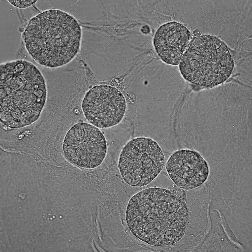
Organoids are a type of 3D culture system. As the word suggests, an organoid is an artificial mini-organ that presents some characteristics similar to the organ of origin. One particular feature of this type of cell culture is that it not only produces cell-cell interactions but also cell-matrix interactions, which are crucial for the architecture and development of organs.
Organoids can be developed from samples of normal tissue, such as the intestine, kidney or the brain. These models help in understanding different processes involved in organ development, regeneration, and disease. Furthermore, organoids can also be obtained from malignant samples, like a tumor.
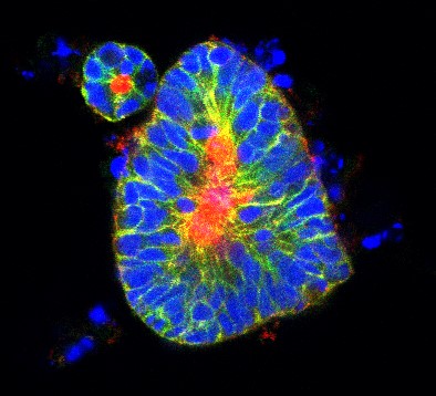
Organoids have emerged as a powerful alternative to traditional animal models, offering scientists a faster, more precise way to explore complex biological questions. These 3D in vitro systems closely mimic the structure and function of real organs, providing unprecedented insights into human development, disease progression, and drug responses. As this field continues to expand, researchers are uncovering new ways to tackle some of the most challenging scientific and medical puzzles, accelerating discoveries and reducing the reliance on animal testing
References
Zhao, Z., Chen, X., Dowbaj, A.M. et al. Organoids. Nat Rev Methods Primers 2, 94 (2022). https://doi.org/10.1038/s43586-022-00174-y
Koo B, Choi B, Park H, Yoon KJ. Past, Present, and Future of Brain Organoid Technology. Mol Cells. 42(9):617-627 (2019) . doi: 10.14348/molcells.2019.0162.
Sato, T., Vries, R., Snippert, H. et al. Single Lgr5 stem cells build crypt-villus structures in vitro without a mesenchymal niche. Nature 459, 262–265 (2009). https://doi.org/10.1038/nature07935